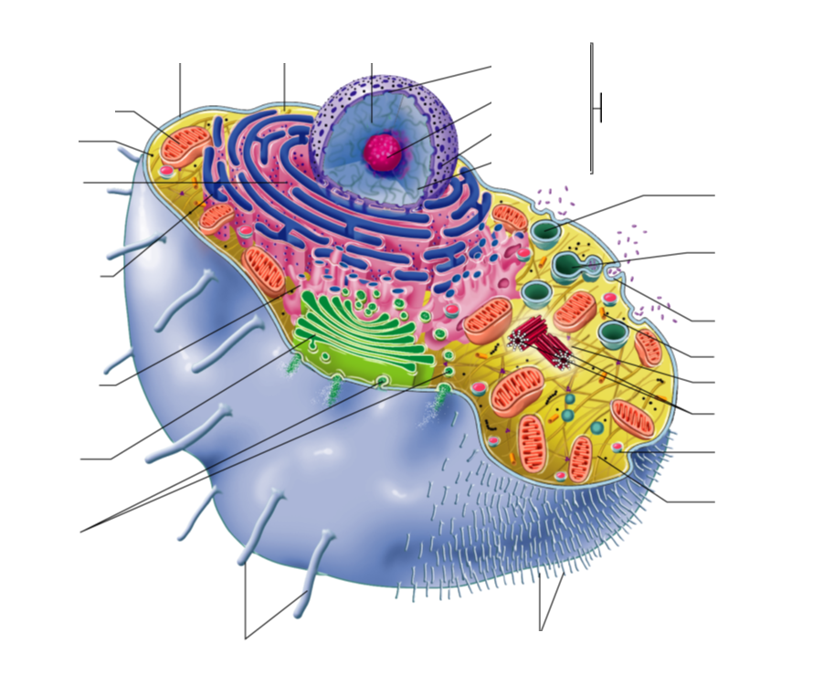
generalized human cell, labeled Diagram Quizlet
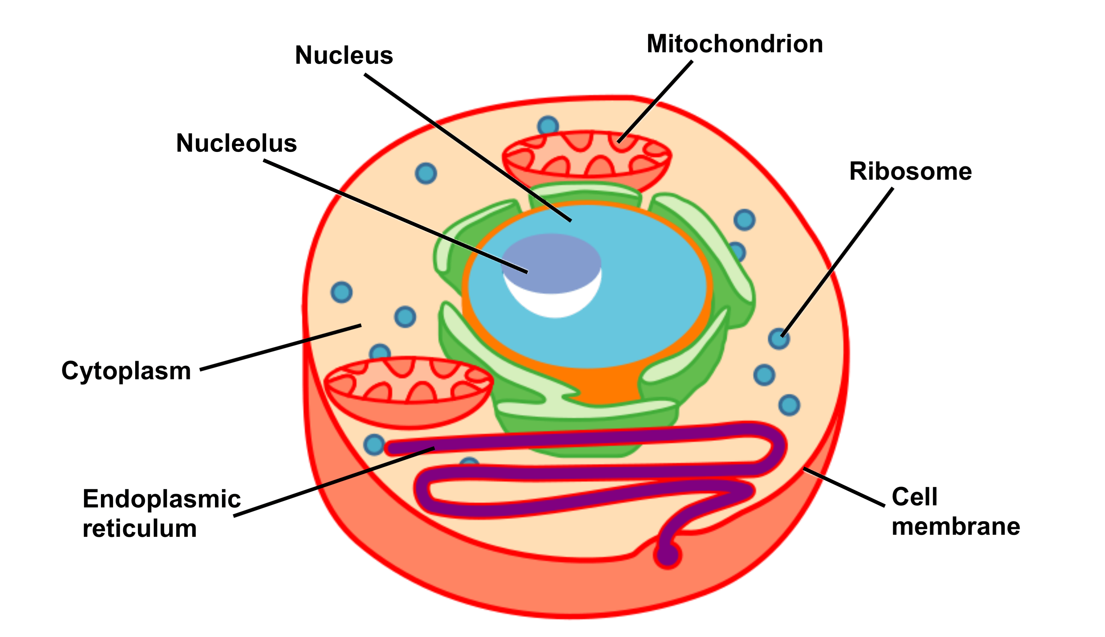
I've created two interactive diagrams for an upcoming open textbook for high-school level biology. The cell structure illustrations for these diagrams were generated in BioRender. Both diagrams feature a drag-and-drop labelling activity created with H5P here on Learnful. These h5p resources are made available openly with the CC BY license.
Structure Of Human Cell With Labels Images & Pictures Becuo
Cell organelles are specialized entities present inside a particular type of cell that performs a specific function. There are various cell organelles, out of which, some are common in most types of cells like cell membranes, nucleus, and cytoplasm. However, some organelles are specific to one particular type of cell-like plastids and cell.
Education Chart of Biology for Human Cell Diagram Best Acupuncture llc
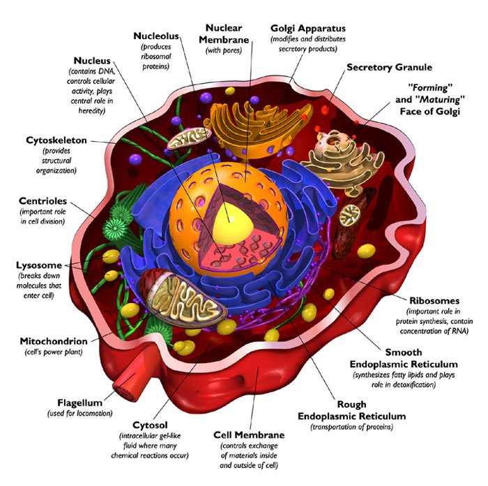
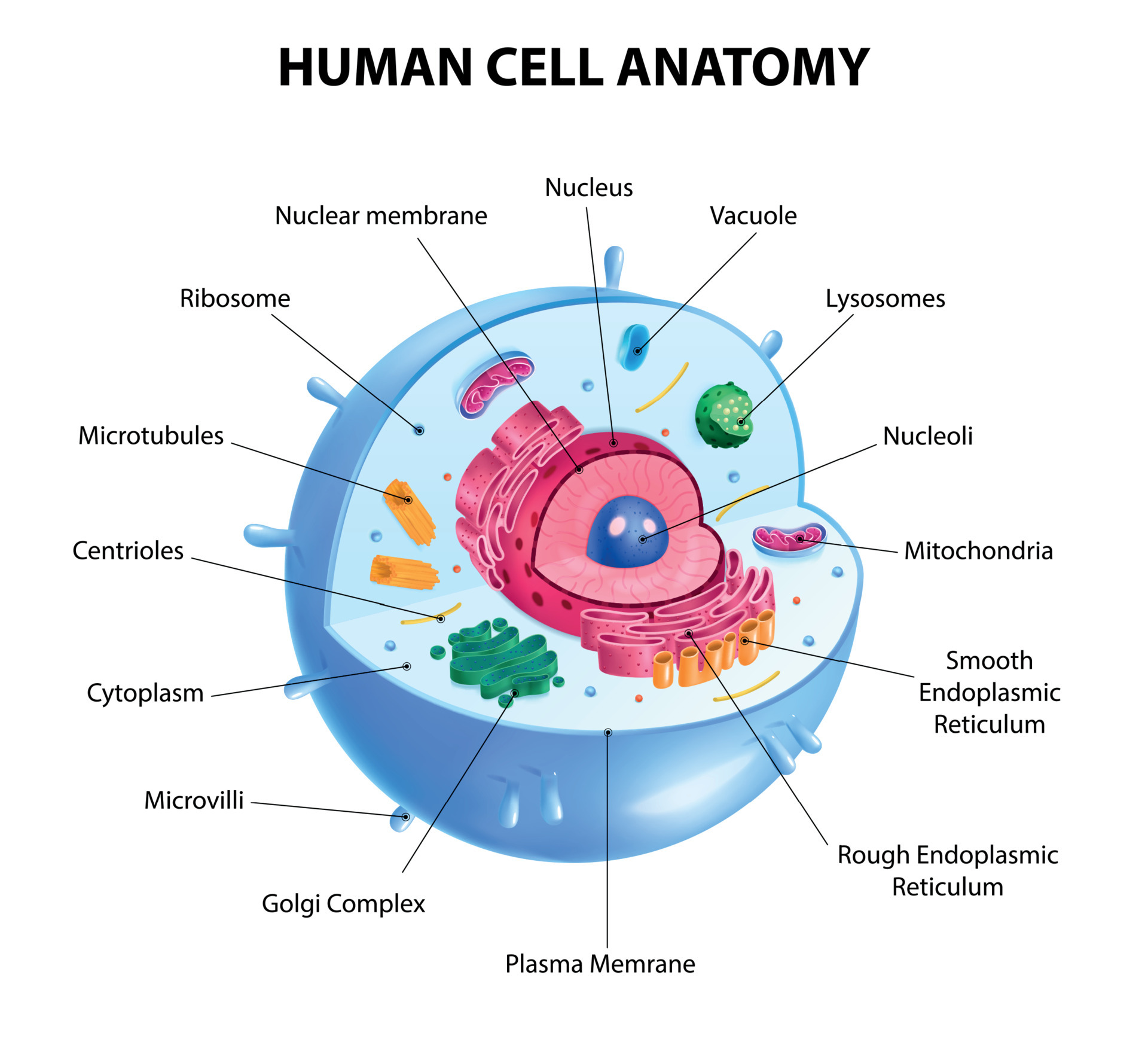
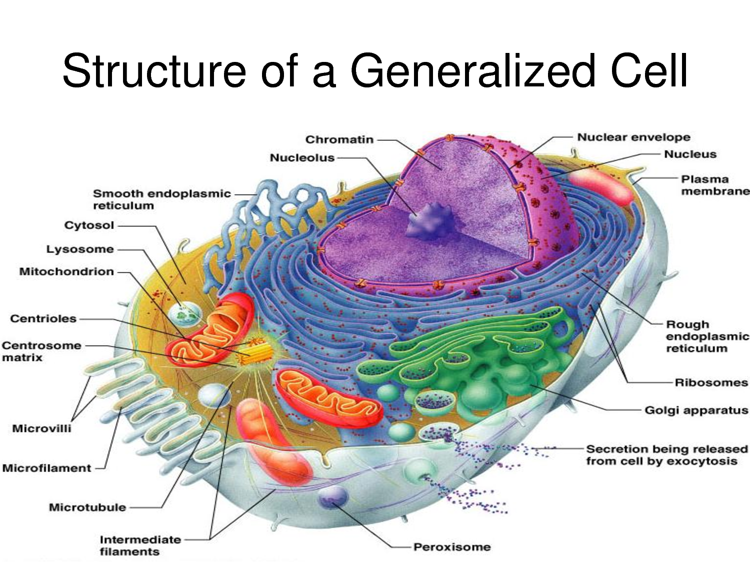
Diagram 1: The anatomical presentation of the human cell. Picture Source: www.printablediagram.com How many cells are in the human body ? Ans : Approx. 37.2 trillion cells What are the different parts of the human cells? How do these parts function? Cell membrane It is the outer covering of the cell, which consists of proteins and lipids.
Cell Membrane Definition Biology Functions Cell Diagram
Interactive guide to stem cells and cell biology with 3D models and real microscopy data of GFP labeled hiPSCs.
Human Cell Diagram 6406474 Vector Art at Vecteezy
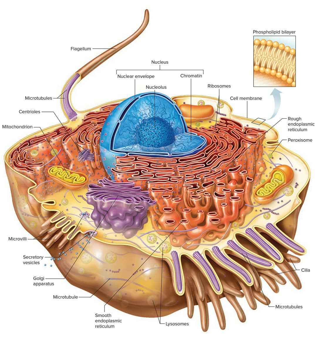
Diagram of the human cell illustrating the different parts of the cell. Cell Membrane The cell membrane is the outer coating of the cell and contains the cytoplasm, substances within it and the organelle. It is a double-layered membrane composed of proteins and lipids.

Cells
3d cell stem science background. Medical microscopic molecular conception. Biology research dna nucleus cells vector pattern. 3D illustration of the internal and external structures of HIV (human immunodeficiency virus). Also seen are antibodies and T helper cells (large spheres with protrusions).
Cell Structure and Function Part 1 The Organelles Medical Exam Prep
Cell (biology) The cell is the basic structural and functional unit of all forms of life. Every cell consists of cytoplasm enclosed within a membrane, and contains many macromolecules such as proteins, DNA and RNA, as well as many small molecules of nutrients and metabolites. [1] The term comes from the Latin word cellula meaning 'small room'.
Human cell hires stock photography and images Alamy
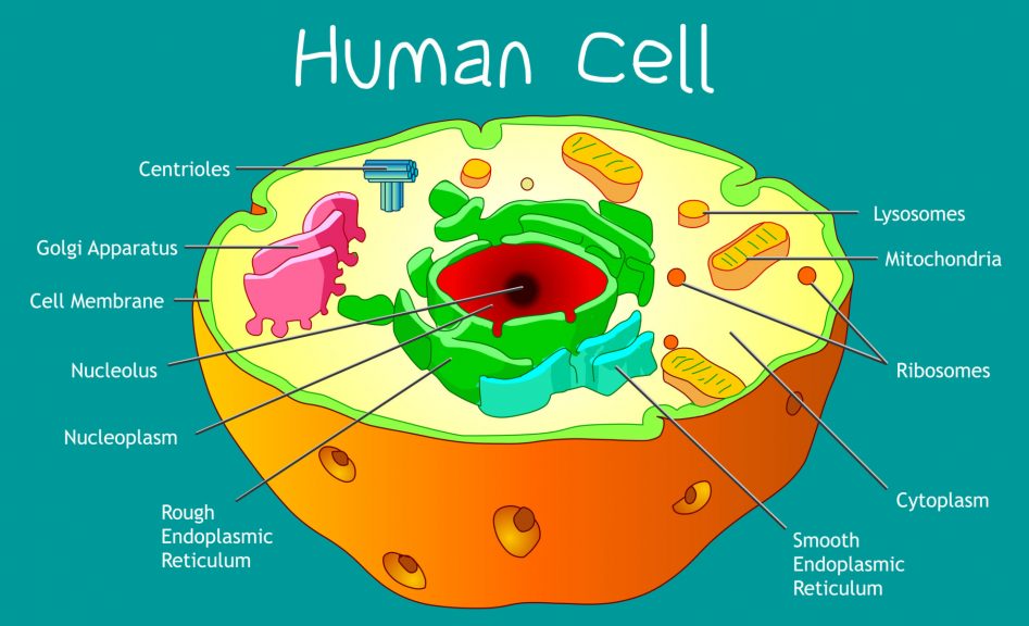
Key points: All cells have a cell membrane that separates the inside and the outside of the cell, and controls what goes in and comes out. The cell membrane surrounds a cell's cytoplasm, which is a jelly-like substance containing the cell's parts. Cells contain parts called organelles. Each organelle carries out a specific function in the cell.

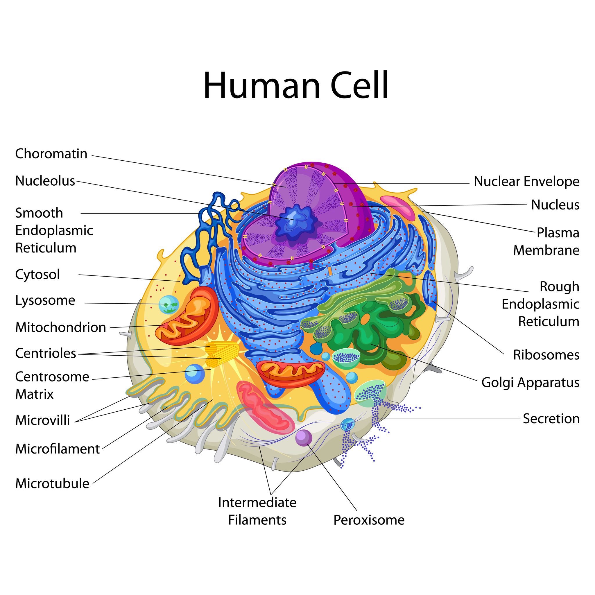
Labeled Diagram Human Cell
What are the parts of a cell? There exist two general classes of cells: Prokaryotic cells: Simple, self-sustaining cells (bacteria and archaea) Eukaryotic cells: Complex, non self-sustaining cells (found in animals, plants, algae and fungi) In this article, we'll be focusing on eukaryotic cells. Two major regions can be found in a cell.
Cells Haleo
Diagram of the human cell illustrating the different parts of the cell. Cell Membrane The cell membrane is the outer coating of the cell and contains the cytoplasm, substances within it and the organelle. It is a double-layered membrane composed of proteins and lipids.

Cell Structure
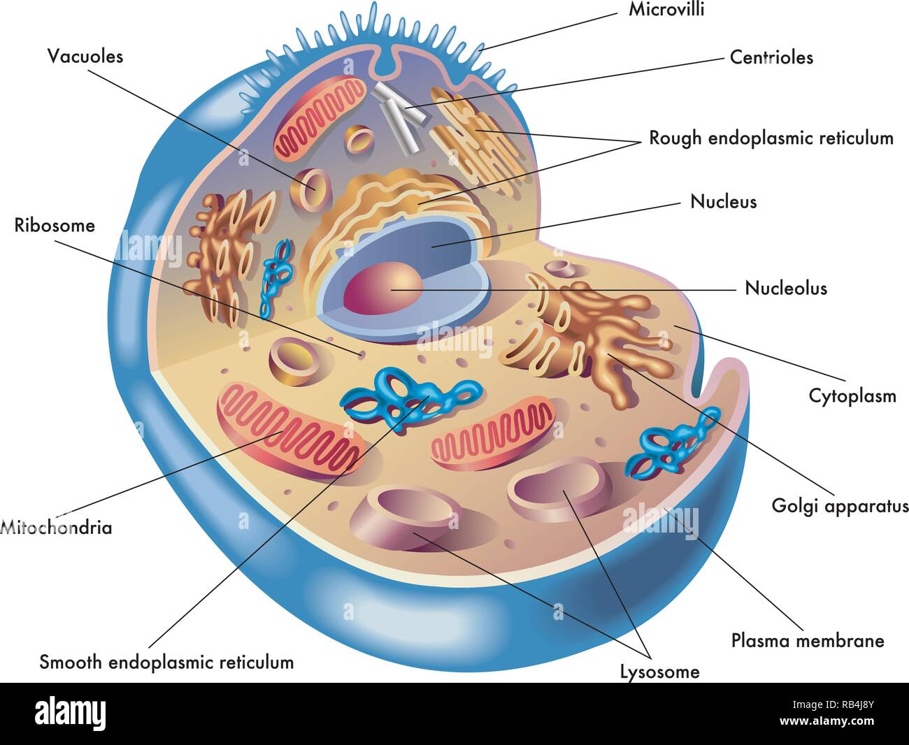
A labelled diagram of a human cell is shown below. Each of the labelled structures will then be discussed individually. Cytoplasm The cytoplasm is the jelly-like substance, that primarily consists of water (approx. 90%) and fills the majority of the cell. In addition to water, the cytoplasm also contains dissolved nutrients and waste products.
Human Cell Diagrams Images & Pictures Becuo
Article Shared by ADVERTISEMENTS: Let us make an in-depth study of the structure and functions of cell. After reading this article you will learn about: 1. Comparison of Prokaryotic Cells and Eukaryotic Cells and 2. Structure and Components of a Human Cell. Cell is a compartment where all the activities of life takes place.
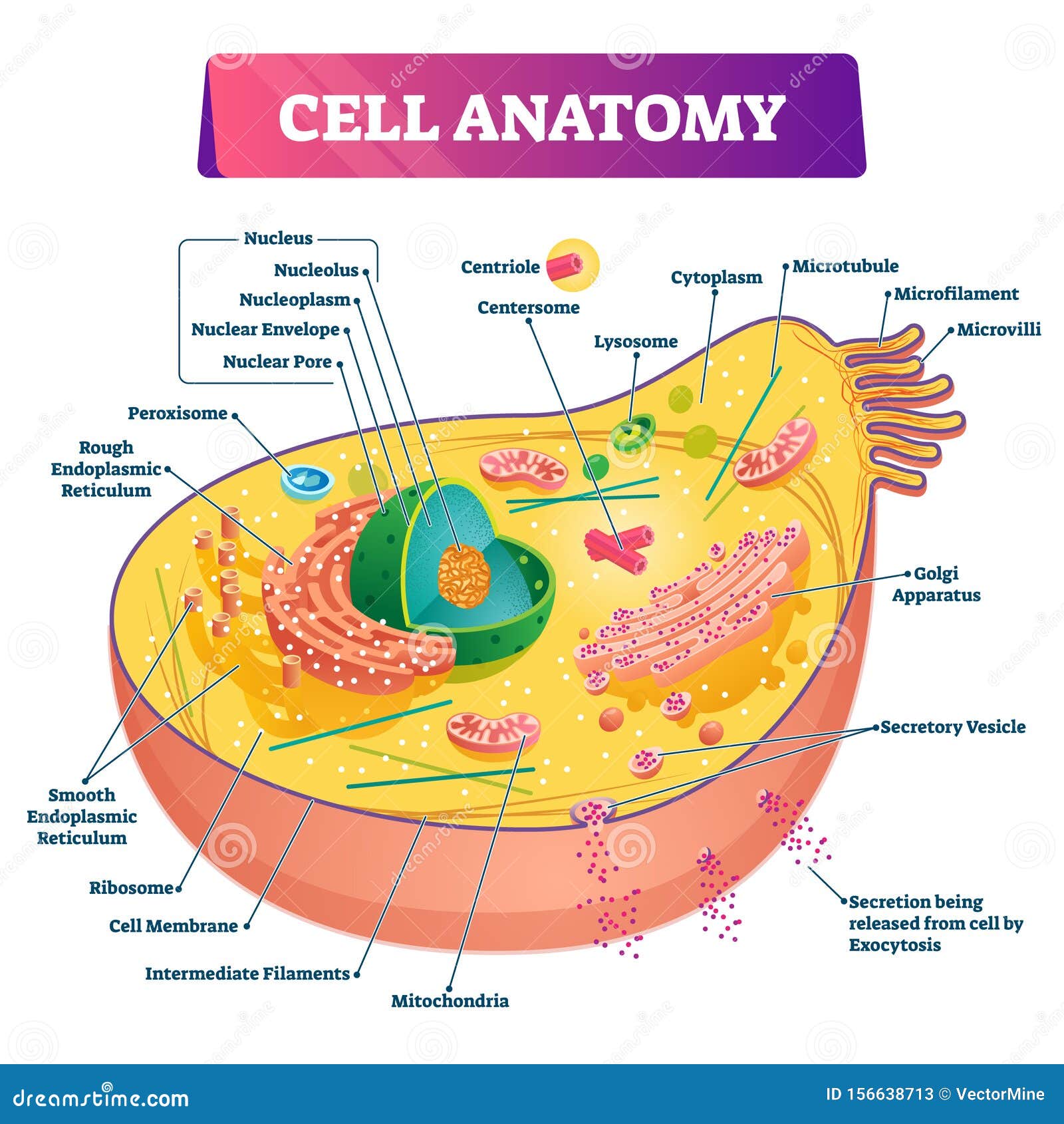
Cell Anatomy Vector Illustration. Labeled Educational Structure Diagram Stock Vector
A cell is the smallest living thing in the human organism, and all living structures in the human body are made of cells. There are hundreds of different types of cells in the human body, which vary in shape (e.g. round, flat, long and thin, short and thick) and size (e.g. small granule cells of the cerebellum in the brain (4 micrometers), up to the huge oocytes (eggs) produced in the female.
Cell Biology, Cell Structure
Human Cells. Part of Human Biology. Human Cells. Division and differentiation in human cells. When cells express specific genes that characterise a certain type of cell we say that a cell has.

69,023 Human Cell Structure Images, Stock Photos & Vectors Shutterstock
The nucleus is a large organelle that contains the cell's genetic information. Most cells have only one nucleus, but some have more than one, and others—like mature red blood cells—don't have one at all. Within the nucleus is a spherical body known as the nucleolus, which contains clusters of protein, DNA, and RNA.

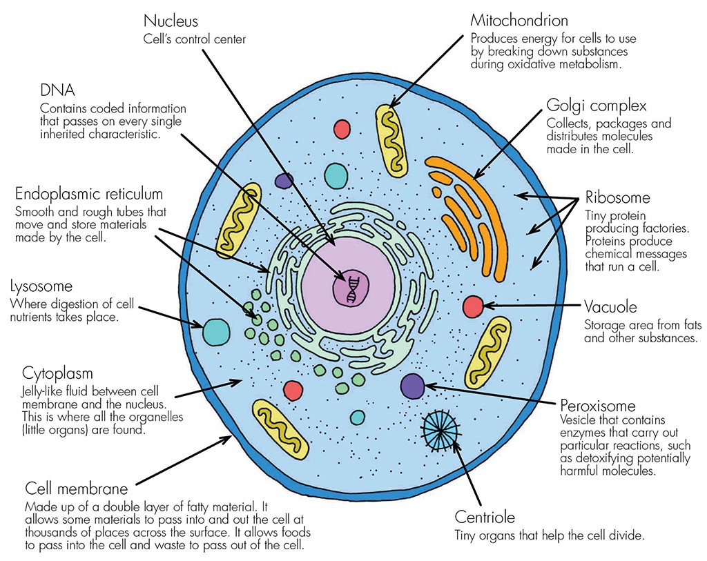
Infographic Anatomy of a Cell
A labeled human cell diagram can highlight important structures such as the cell membrane, nucleus, cytoplasm, mitochondria, endoplasmic reticulum, Golgi apparatus, and more. By referring to such a diagram, individuals can better understand the function and organization of these cellular structures.
